
GE Healthcare Debuts Next-Generation Nuclear Medicine Cardiology System
GE Healthcare recently unveiled MyoSPECT, a next-generation cardiac-dedicated nuclear scanner with extended field-of-view processing and new automated workflow features for a fast, comfortable exam experience.
Mon Oct 04 2021
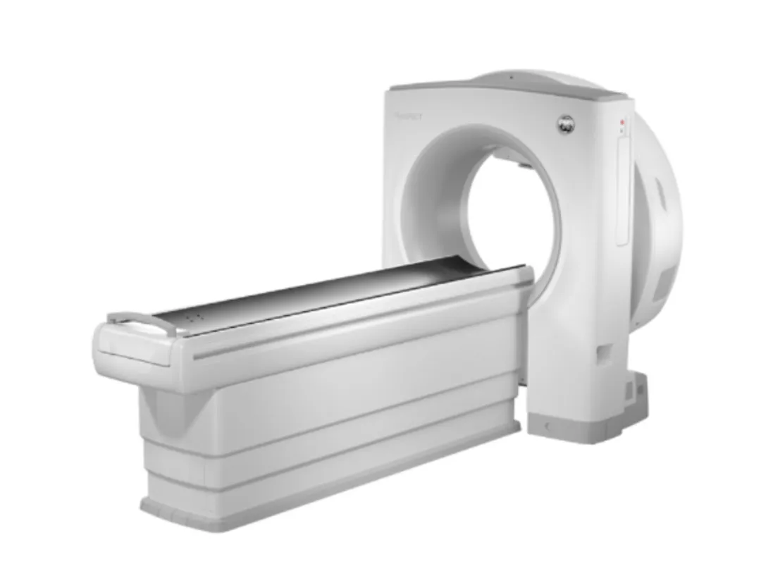 Built with the company’s exclusive Alcyone technology and CZT module design, MyoSPECT offers an exceptional view of cardiac anatomy and pathology to help clinicians determine the right course of treatment for patients.
As the number of people diagnosed with and dying from cardiovascular disease (CVD) steadily increases[iii], so too does the number of cardiac procedures, which now account for over 50 percent of all nuclear medicine studies in the United States[iv]. This evolving situation underlines the need for new cardiac-specific imaging solutions that deliver accurate assessments for patients of all sizes.
“Today, the need for healthcare innovation is stronger than ever,” explains Jean-Luc Procaccini, President & CEO, Molecular Imaging & Computed Tomography, GE Healthcare. “Not only is demand for nuclear cardiac solutions increasing, but new and tough cardiac cases require a different approach to patient comfort, image quality, and workflow. That’s why we designed MyoSPECT with all these factors in mind. Not only does MyoSPECT offer extended field of view processing to help accommodate a greater variety of patients, but its digital CZT detectors and new automated workflow features also help expedite exams for fast, confident diagnoses.”
MyoSPECT offers a wider table and 76 percent increase in field-of-view volumei, providing clinicians greater positioning flexibility to help accommodate more patients, including those who may be too weak to sit up through an entire exam or others with high body mass indexes (BMIs). Combined with the excellent energy and spatial resolution of GE Healthcare’s compact CZT detector technology and novel multi-pinhole collimator design, these features create a tomographic imaging arc of the heart with motionless detectors, so every detector focuses on the heart simultaneously. The result is a comfortable experience for patients of all sizes and a complete picture of the heart’s anatomy and pathology for clinicians.
“From form to function, there has never been a more complete solution for cardiac imaging,” says Prof. Tali Sharir, director of nuclear cardiology at Assuta Medical Centers in Tel Aviv, Israel – the first evaluation site for MyoSPECT. “MyoSPECT provides very good image quality for obese patients with short scanning times, easy workflow, and excellent patient comfort. Also, the system’s new capabilities – including Smart Positioning, extended field-of-view processing, and imaging protocol – helped us expedite our department’s typical two-day protocol to a one-day protocol instead.”
Recognizing that many healthcare systems today are challenged to maintain quality across a wide range of patient and exam types, MyoSPECT also includes several new automated features to help expedite workflows and make it easy to achieve quality results consistently.
To start, the system’s new Smart Positioning solution offers scan positioning prompts and extended field-of-view recommendations to optimize patient positioning. Then, MyoSPECT leverages device-less Alcyone Motion Detection and Correction on Xeleris – GE Healthcare’s processing and review solution – to generate images without motion to view cardiac anatomy and pathology with fewer artifacts and great clarity. The system also includes SPECT Flow, which combines dynamic acquisition on MyoSPECT with coronary flow reserve (CFR) and absolute myocardial blood flow, should clinicians wish to use an external CT for attenuation correction[v]. This process delivers valuable insight into each patient’s blood flow for true dynamic cardiac imaging and great diagnostic confidence.
Finally, the system offers clinicians two options for attenuation correction and evaluation – both of which have the added benefit of excellent CZT resolution:
Correct attenuation by combining perfusion images with separate CT images on Xeleris; or
Evaluate attenuation artifacts by imaging both prone and supine positions without additional radiation exposure since MyoSPECT uses a table for patient positioning.
Altogether, the system is designed to view cardiac anatomy and pathology with great clarity, exceptional patient comfort, and provide clinicians with the information they need to help improve patient outcomes.