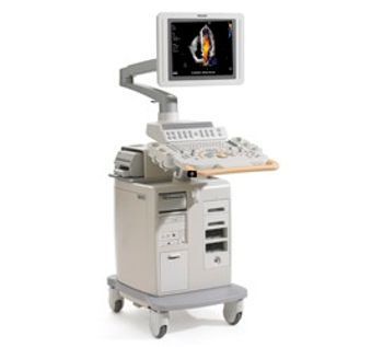
Philips - HD11 XE
by Philips


Ultrasound System
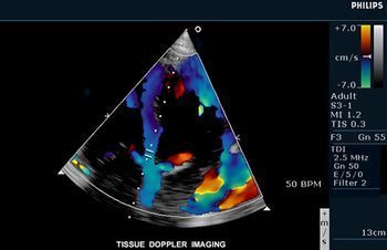
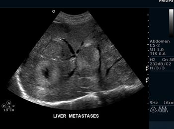
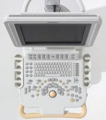
In this full-featured performer, Philips has combined broadband beamforming, automated image optimization tools, and clinically proven technologies. The HD11 XE is ideal for public and private hospitals, satellite clinics or specialized practices. And it’s built on an upgradeable platform to protect investments, backed by Philip’s customer support program. Includes SonoCT spatial compounding image acquisition and XRES adaptive image processing. Also 4D imaging, qualitative freehand 3D, 2D pulse inversion harmonic imaging, and QLAB quantification software.
0Replies4 hours ago | Image area size smaller than normal The image area seems smaller than before. I assume I have changed a setting but I can't get it back to normal size. Does somebody has suggestions? |
0Replies18 hours ago | Window is smaller as usual Hello, suddenly my screen became a bit smaller than usual. The menu is full screen only the image seems smaller than before. It looks like the window is based on 15" instead of 17". It was good but I might have select some settings. How to turn back to normal? 2nd question: can somebody provide a service manual for the HD11 XE? I have the manual of HD11 but this one seems to be different.. Thanks in advance! Equipment: Philips - HD11 XE Reply |
1Reply7 months ago | message d'erreur interne Bonjour, Mon appareille d'écho affiche ce message d'erreur : "L'instrument a détecté une erreur interne et collecte des informations pour aider à diagnostiquer le problème / Veuillez contacter votre représentant Philips. La commande "Redémarrer" sera activée lorsque les informations auront été obtenues. . " Veuillez conseiller sur la façon de le résoudre. Merci. Equipment: Philips - HD11 XE Reply |
| Cart Based | 1 |
| Clinical Applications | Colorectal |
| Doppler Modes | 3-D |