
OrthoScan - UC
by OrthoScan
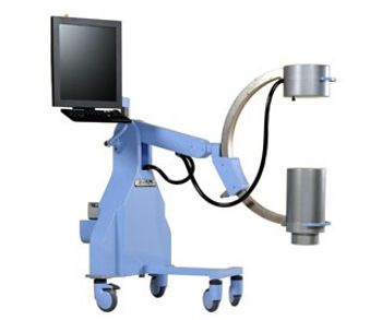
Mobile C-Arm
OrthoScan UC Mini C-Arm is 1/2 the size and 1/3 the weight of the competition. The smaller size facilitates ease of movement and improves maneuverability. OrthoScan is putting control back in the surgeon’s hands and making a difference in the experience for every user. OrthoScan UC brings the surgery closer to you. The superior display size of the image allows for more precise evaluation of images taken and enables users to analyze fine details of the image at a distance. The “live” image is 28% larger than our competition’s largest image. Just like viewing a big screen TV from across a living room, it is easier to see a larger image at a distance. The resolution of the OrthoScan UC enables the highest quality of imaging detection through superior gray scale and exceptional clarity. The improved diagnostic quality provides the ability to detect hairline fractures that may have been overlooked. OrthoScan UC automatically adjusts to metal, motion, and varying anatomy. The accurate adjustments of the OrthoScan UC Mini C-Arm allow the operator to use less fluoro time (less dose) and accurately view anatomy. It allows the surgeon to focus on the patient rather than equipment. OrthoScan UC has a simple interface that allows the user to perform basic imaging with little, if any, preparation. The custom keyboard allows single key access to most functions. The features are accessible, intuitive, and – for the most part – automatic.
4RepliesFri Feb 22 2019 | Service Manual Hi I am in need of a service manualReply |
4RepliesTue Sep 27 2016 | DiCOM
I am unable to connect to DiCOM. A message appears when I try to send an image that say "unable to initialize DiCom store" Is this an ethernet card failure? Reply |
8RepliesFri Mar 21 2014 | Service Manual/PM Due I am in need of a service manual Reply |
| Image Display | 18.1 in or 20.1” (with optional monitor upgrade) |