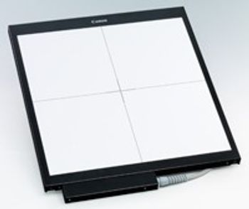
Canon - Canon CXDI-40G Compact Digital Radiography System
by Canon


Digital Detectors
EXPANDING THE BENEFITS OF DIGITAL RADIOGRAPHY The CXDI-40G COMPACT is equipped with the large image sensor of 43 cm x 43 cm and high-quality capabilities of the CXDI-40EG but in minimal housing. This can be easily retrofitted into a range of radiography devices such as upright stands, RF tables and Bucky units.
Upgrade your current equipment with CXDI-40G COMPACT and greatly streamline radiographic workflow. Results in Seconds an on screen preview image is available in just 3 seconds after exposure. If another exposure is required, the detector will be ready in seconds thanks to its fast refresh cycle. MLT(S) Image Processing MLT(S) image processing utilizes multi frequencies to emphasize on details and edges. In combination with the appropriate dynamic range and Canon’s advanced noise reduction processing images show subtle details like lung tissue or trabecular bone structure together with soft tissue in high diagnostic accuracy. These parameters can be preset in our dedicated body parts for repeatability and adjusted post-acquisition for variances in diagnostic requirements
Canon’s Lanmit Technology Amorphous Silicon (a-Si) Flat Panel Sensor (LANMIT) consists of a precision amorphous silicon metal-insulator semiconductor (MIS) sensor and Thin Film Transistor (TFT) array. LANMIT flat panel sensors produce a highly stable output signal. The result is a family of reliable and durable sensors that provide the highest quality images without the time and costs involved with film or cassette handling. A 14-bit analog-to-digital conversion process converts the raw image to digital form producing a high-resolution, high-contrast 4,096 grayscale image. Seamless Network Integration Ethernet 10/100/1000 Base T connectivity with DICOM 3.0 compatibility enables seamless data transfer to any DICOM hard copy output device, PACS, or HIS/RIS for efficient printing, archiving, and remote viewing of images. The system complies with IHE (Integrating the Healthcare Enterprise) standards for interoperability to ensure easy, effective integration with the hospital network.
| Weight | 24 lbs 11kg, (without cable) |